Osteoarthritis and the Impact of Obesity
Obesity is strongly associated with osteoarthritis, and in particular a dose dependent relationship has been found to exist between BMI and risk of developing osteoarthritis of the knees. Excessive osteoarthritis of otherwise healthy joints is known to contribute towards osteoarthritis, but obesity is also a risk factor for developing osteoarthritis in non-weight-bearing joints, and current research indicates a more complex relationship exists between osteoarthritis and obesity than can be explained by biomechanical stress alone.
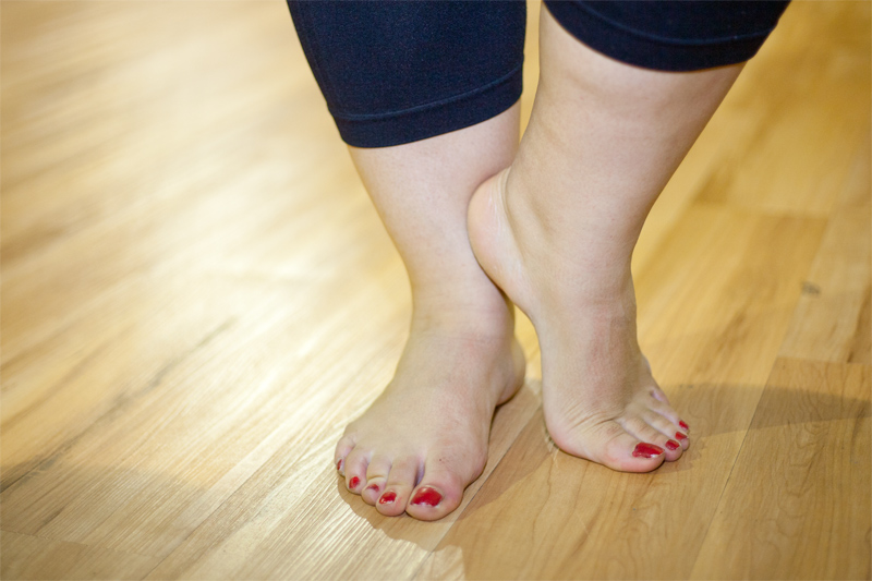
Protect your ankles
What is Osteoarthritis?
Osteoarthritis is a common degenerative condition affecting the joints and surrounding tissues. Developing gradually over time, it most frequently occurs in the hips, knees, cervical and lumbosacral spine, base of the big toe and small joints of the hands, but can affect almost any joint in the body.
The Knee

The bones of a joint are held together by ligaments that keep them in place whilst the joint is in motion. The surfaces of each bone are costeoarthritisted in a layer of articular cartilage which serves to distribute loading across the joint, protect the bone surfaces and reduce friction. A highly vascular layer of bone, known as subchondral bone is located directly below the cartilage. The joint itself is housed within a capsule and the joint cavity within is filled with synovial fluid generated by a layer of synovial membrane lining the cavity. Synovial fluid delivers essential nutrients to the joint and avascular articular cartilage, and also functions as a shock absorber.
In general terms osteoarthritis occurs as a result of articular cartilage degeneration. As the cartilage breaks down, the underlying subchondral bone starts to thicken and brosteoarthritisden in an attempt to reduce losteoarthritisd on the cartilage, forming bony growths called osteophytes at the joint margins. The synovial membrane and capsule thicken, reducing space inside the joint, and in the early stages of the condition pathologic changes in the synovial fluid can cause the cartilage to swell. Over time, further chemical changes cause the cartilage to soften and lose elasticity, eventually eroding to the extent that the underlying bone is exposed. This causes trauma to the bone, puts strain on the ligaments and can lead to the formation of osteosteoarthritisrthritic cysts. Nerve damage and atrophy of the surrounding muscle may also occur.
What are Risk Factors for Osteoarthritis?
Pain during movement and swelling of the joints are common symptoms. Long periods of inactivity such as during sleep can cause stiffness. If the cartilage cover is totally worn out then the pain is present even when at rest. The symptoms vary with the patient; some patients may report little pain or discomfort even with very obvious degeneration of the joints. The signs and symptoms can appear sporadically. Afflicted patients can experience prolonged periods of pain-free existence. Symptoms will also vary with the joint being afflicted.
Osteoarthritis is classified as into primary or secondary condition depending upon the detection of an underlying cause. With age, the cartilage begins to degenerate and retain water. The cushioning effect of the cartilage reduces and the joints are exposed to wear. Friction between the cartilage and joint can lead to swelling and pain in the joints. Joint movement becomes restricted. Sometimes, osteoarthritis is characterized by growth of bony spurs around the afflicted joints. There is evidence to suggest that primary osteoarthritis is often genetic and hereditary.
Osteoarthritis may be classified as secondary to an identifiable cause such as trauma or congenital joint deformity, or primary, when no underlying cause can be determined. Cartilage naturally loses its resilience as part of the ageing process, and primary osteoarthritis is most common in those over 50 years of age, with the majority of people aged over 70 years showing signs of osteoarthritis in at least one joint. However, not all of those individuals will be symptomatic, and osteoarthritis is not an inevitable consequence of age.
Cases of osteoarthritis in those aged under 40 years display a male preponderance and are frequently precipitated by traumatic injury to the affected joint. Beyond 50 years of age, female cases predominate, possibly as a result of hormonal effects.
Genetic factors are thought to play a significant role in the pathogenesis of osteoarthritis, and various genes have been identified as contributing to overall osteoarthritis susceptibility and influencing specific manifestations of the condition. Congenital issues directly affecting the joint are also a known risk factor, the extent of which is difficult to quantify as even very subtle defects may be sufficient to initiate osteoarthritis.
Secondary osteoarthritis may be associated with metabolic disorders such as diabetes and Wilson’s disease, connective tissue disorders including Marfan Syndrome and Ehlers-Danlos Syndrome and inflammatory diseases such as Lyme disease and Perthes’disease. Additionally, osteoarthritis may develop secondary to joint damage caused by other forms of arthritis, most notably Rheumatoid arthritis.
Signs and Symptoms of Osteoarthritis
Diagnosis is made on the basis of clinical presentation and radiographic findings, with lab tests performed to rule out differential diagnoses including Lyme disease, joint infection or Rheumatoid arthritis, or to investigate a possible underlying cause of secondary osteoarthritis.
The Feet

Externally, swelling may be apparent, and osteophytes may lead to visible enlargement, particularly in the smaller joints such as those of the hands and feet. When affecting the distal (closest to the fingertip) or interphalangeal (middle) joints of the fingers they are known as Heberden’s nodes and Bouchard’s nodes respectively. When the feet are affected, bunions may form. Medical imaging may reveal increased subchondral bone density, subchondral cyst formation, osteophytes, effusion and reduced space within the joint.
Whilst it is possible to have asymptomatic osteoarthritis, joint pain that is exacerbated by use is often the main presenting symptom. The range of motion of the affected joint may become restricted, and prolonged lack of movement during periods of rest or sleep can lead to joint stiffness or “gelling” that may persist for up to 30 minutes. Joint effusion or swelling is also common, although not necessarily accompanied by pain or restriction of movement, and osteoarthritis is a significant cause of effusion of the knee joint, otherwise known as “water on the knee”. Some people also experience crepitus; a cracking sound that may be accompanied by a grinding or crunching sensation when the joint is in motion.
What Joints are Commonly Afflicted by Osteoarthritis
- Base of the thumb – Commonly found in post-menopausal women. The exact cause is not known; however, heredity and a previous fracture can lead to the condition.
- Knees – Osteoarthritis of the knee joints is often the result of age, ligament injury, or fracture. The condition is common in obese individuals.
- Wrist – The condition can develop over time from the wear and tear that the wrist joint and cartilage suffer. Injury to the wrist or forearm can also cause osteoarthritis.
- Shoulder – Osteoarthritis of the shoulder is usually found in people above 50 years of age.
- Hip – As with the other weight-bearing joint of the hip, here too obesity is a cause. Wear and tear of the joint over a period of time can also cause arthritis.
What Tests are Used for a Diagnosis of Osteoarthritis
X-rays are a possible diagnostic test for the condition. Abnormalities that show up include loss of cartilage covering the joint and bone spurs. Blood tests are performed to check for diseases that can cause secondary osteoarthritis. X-rays are also helpful in excluding other probable causes and determining the best treatment option. MRI and CT-Scan are other imaging techniques used.
A procedure called arthocentesis is sometimes used to exclude gout and infection as causes of the condition. It involves removal of joint fluid via a sterile needle and analyzing the fluid.
Arthroscopy is used to view the joint spaces using a tube inserted there. It enables the medical practioner to check for damaged cartilage and lax ligaments.
Physical examination, the presence of Heberden's nodes and bunions on the feet, questioning on the nature and duration of symptoms can help doctors in diagnosing osteoarthritis.
How does Obesity affect Osteoarthritis?
Adipose tissue, particularly when localized to the abdominal area, is known to be metabolically active, producing various factors including the hormones leptin, adiponectin and resistin. These chemical messengers can mediate changes in bone growth and cartilage homeostasis, and leptin in particular has been found to exist in elevated concentrations relative to blood plasma within the synovial fluid of osteoarthritis joints. Furthermore, laboratory tests on cell and tissue culture models have shown the presence of leptin to be associated with increased cartilage degradation.
Vascular changes in the subchondral bone have been observed in osteoarthritis joints and cardiovascular disease is a known risk factor for osteoarthritis. Hence it has been hypothesized that restrictions in blood supply may have a direct ischemic effect upon the bone, and/or impact upon the supply of nutrients to the cartilage, either of which could potentially accelerate the rate of progression of osteoarthritis. Cardiovascular disease and metabolic disorders such as diabetes are strongly associated with obesity and known to be independent risk factors for osteoarthritis, even when controlling for BMI, suggesting that a common set of metabolic factors may be implicated in the pathogenesis of these conditions.
Whilst wear and tear in the form of static joint loading plays a role in the initiation and progression of osteoarthritis, some forms of exercise have been shown to have a protective effect against cartilage degeneration. Exercise is generally considered to have an anti-inflammatory effect, and cyclic joint loading may positively influence joint health through direct stimulation of the articular cartilage, which can inhibit inflammation and protect against cartilage degeneration. Research indicates that a significant number of obese individuals with osteoarthritis have a sedentary lifestyle, suggesting that lack of regular exercise may contribute to development of osteoarthritis in such cases, possibly as a result of increased susceptibility to inflammation.
Obesity related Osteoarthritis and Exercise
Can Bariatric Surgery help Osteoarthritis?
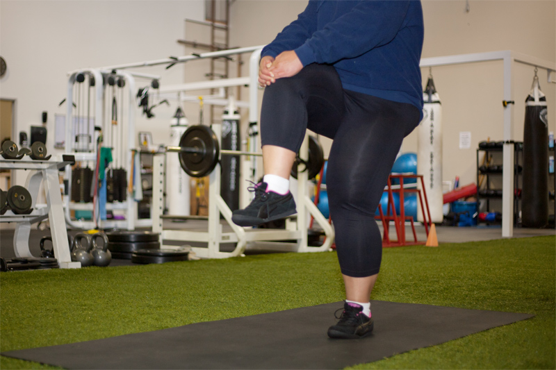
In overweight and obese individuals, weight loss can simultaneously reduce the degree of harmful static loading upon weight-bearing joints and increase overall mobility, making it easier to participate in regular exercise. Additionally, the positive effects of weight loss upon cardiovascular and metabolic status may reduce overall susceptibility to osteoarthritis. However, a combination of obesity and osteoarthritis can make exercise extremely difficult, exacerbating osteoarthritis symptoms and preventing weight loss. In such cases, the rapid weight loss of fifty to one hundred pounds typically facilitated by bariatric surgery can provide an effective means of breaking that cycle, with loss of weight providing immediate relief from static loading whilst simultaneously improving mobility and exercise tolerance such that a program of regular exercise and physical therapy designed to offer longer term improvements in osteoarthritis symptoms can be undertaken.
What are Other Treatments for Osteoarthritis?
Regular exercise and physical therapy techniques are frequently effective in managing symptomatic osteoarthritis, and in some cases may even halt or reverse osteoarthritis of the hips and knees. An osteoarthritis exercise program should include a combination of activities designed to develop overall fitness, strengthen muscles and increase flexibility. Incorporating regular but brief rest periods into a daily routine can be beneficial, but only in conjunction with exercise and regular usage. Prolonged immobilization of an osteoarthritis joint can result in a condition known as contracture where the soft tissues surrounding the joint become permanently shortened, leaving the joint locked in a flexed position.
In cases of spinal, hip or knee osteoarthritis, avoidance of soft mattresses and deep chairs that encourage poor posture and from which it may be difficult to rise may be recommended, and assistive devices such as supports, leg braces or shock-absorbing footwear may help to reduce pain and increase mobility. Some people obtain symptomatic relief from direct application of a hot or cold pack to the affected area, and performing exercises in warm water can also ease pain whilst helping to prevent contractures.
If analgesia is required, over-the-counter drugs such as Tylenol or Advil may be sufficient to manage pain. Otherwise, stronger painkillers such as non-steroidal anti-inflammatory drugs or NSAIDs may be prescribed. These drugs can simultaneously ease pain and reduce inflammation but are not suitable for everyone, particularly those with a history of asthma, heart attack or stroke. Additionally, when taken regularly they can break down the lining of the gastrointestinal tract, making it susceptible to irritation and damage from stomach acid. Your stomach may be particularly sensitive to NSAID medications after bariatric surgery therefore you need to be sure to discuss these medications with your doctor. Opioids such as codeine are effective in relieving severe osteoarthritis pain, but can cause side effects including nausea, drowsiness and constipation. Topical treatments such as those incorporating NSAIDs or capsaicin (derived from chilli peppers) can help manage the symptoms of osteoarthritis of the hands and knees.
If painkillers are ineffective or contraindicated then a course of intra-articular corticosteroid injections may be recommended. A single dose injected directly into the affected joint can relieve pain, reduce inflammation and increase flexibility for weeks or even months at a time. However, a course of treatment may be limited to three injections per joint per year due to concerns about cartilage damage secondary to treatment.
In a very small number of cases, when conservative treatment methods are ineffective, surgery may be indicated. Minimally invasive arthroscopic procedures can be used to repair or replace damaged cartilage and are particularly effective in treating cartilage tears. Arthroplasty, or total joint replacement is a more invasive procedure involving removal of the joint surface and replacement with a prosthesis. Osteomy, in which small sections of bone are added or removed from the joint in order to re-focus weight away from the most damaged area may be recommended for younger individuals with osteoarthritis of the knee. If no other surgical option is suitable then a procedure known as arthrodensis, in which the joint is fused into a permanent position may be suggested. Whilst this may result in significant pain reduction and a stronger joint, the joint itself will be rendered immobile.
There has previously been some debate over the impact of obesity upon the likely success rate of joint replacement surgery, but recent studies have indicated that arthroplasty procedures such as total knee replacement surgery can facilitate increased mobility in obese individuals, simultaneously promoting sustained weight loss and direct improvement in osteoarthritis symptoms.


