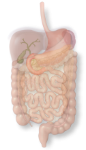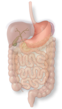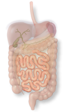

The gallbladder concentrates and stores bile as a pear-shaped sac which it can release to help digestion after a fatty meal.

Laparoscopic tools have opened up opportunities for doctors to take a look inside without a largely invasive procedure. Using these methods, chances of infection are reduces, healing can occur more quickly and the body undergoes less stress. The surgical instruments are introduced into the body through small incisions, along with the camera which is connected to a monitor. The camera allows for a whole different view of the surgical field without opening a large surface on the body.
The magnified view of the surgical field makes finding small bleeders and anomalies easier. It also opens up the possibility of recording video from the camera or capturing still photos which are also helpful to medical students who are getting introduced into the world of surgery. Many interesting shots have been collected from interesting encounters with a variety of cases.
English
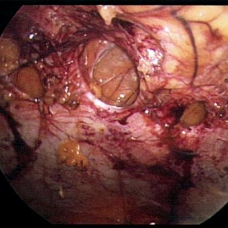
Hernias can become symptomatic because of the altered mechanics of the abdominal wall or because the intestine can become trapped inside of the defects. Multiple defects in are shown in this case which trapped loops of intestine and caused a small bowel obstruction.
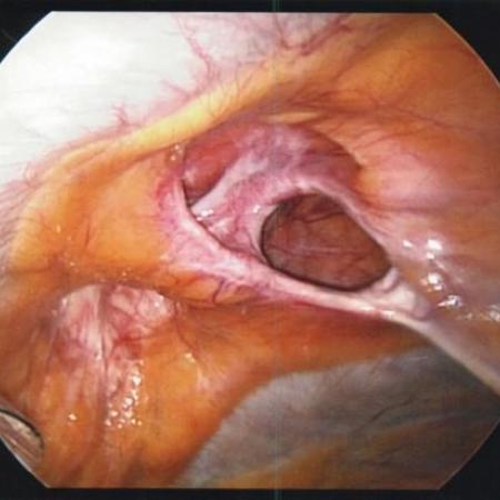
There are three main types of groin hernias, all of which can usually be fixed laparoscopically. These hernias are direct, indirect and femoral and classified according to where they occur in relation to surrounding structures. From an interior view looking down into right groin, a direct space…
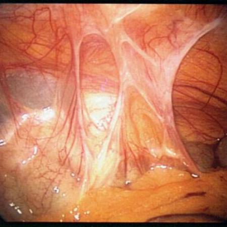
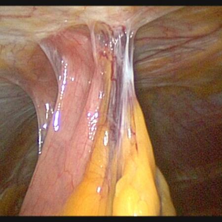
Adhesions between the intestine and the abdominal wall can sometimes develop even if no previous surgery has been performed. Sometimes these adhesions are because of chronic inflammation of the intestine, sometime an exact cause is not discovered. This photo demonstrates an anatomic variant with…
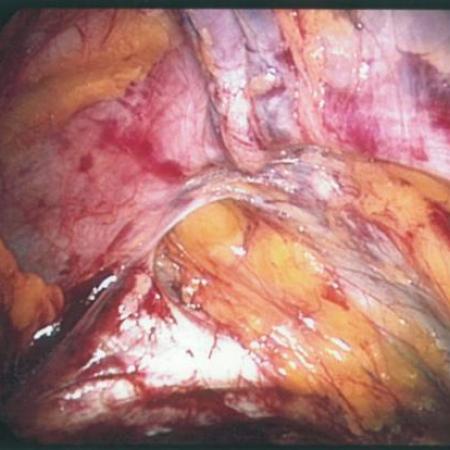
Abdominal wall hernias can be caused from previous operations. The hernia is actually a hole in the muscle. This photo demonstrates a laparoscopic view from inside the abdomen looking up at the multiple holes in the abdominal wall muscle. In this case, multiple large hernias as well as smaller…
Abdominal hernias can sometimes grow very large and contain intestines. The danger of a hernia is that the intestines can become obstructed or the blood supply can become compromised. This view is a laparoscopic perspective showing a loop of colon with small bowel herniating through mesentery into…
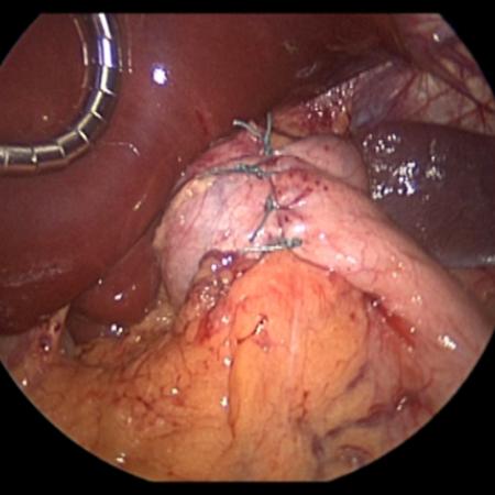
Close-Up view of a Nissen Fundoplication performed for chronic reflux disease that was not alleviated by typical medical management. The gastroesophageal junction is rebuilt during the procedure by wrapping a portion of the stomach, the fundus, around the back of the stomach forming a ring.
Small bowel is shown adherent to the anterior abdominal wall. Scar tissue from a previous open umbilical hernia caused this condition. There is no reliable way to gauge how many adhesions typically exist. The extent of these adhesions can only be seen during an operation.
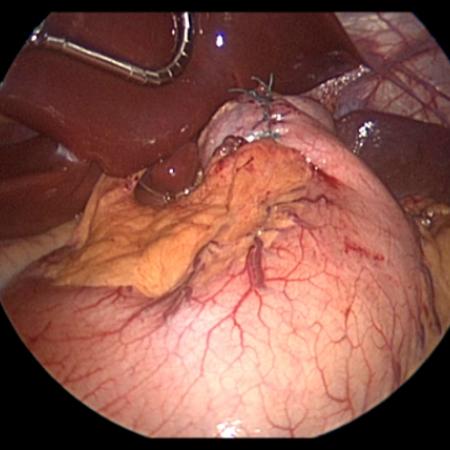
A global view of the stomach after Nissen fundoplication is shown. The liver is the dark pink structure with the metal retractor at the top left of the image. There are four sutures that plicate the top of the stomach wrapping it around the distal esophagus. This plication of the fundus or…
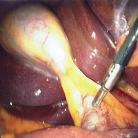
The appendix is a finger-like structure that is attached to the colon. There are conditions when something can block the base of the appendix causing the appendix to become inflamed and infected. An inflamed appendix is shown during a laparoscopic appendectomy. The appendix is in the center of the…
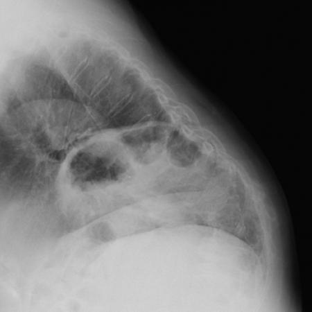
When X-rays of the chest are typically ordered, a front and a side view are typically performed. These views are typically a PA and a lateral view that provide different perspective. This image demonstrates a type IV hiatal hernia with incarcerated colon that was not entirely obvious on the PA view…
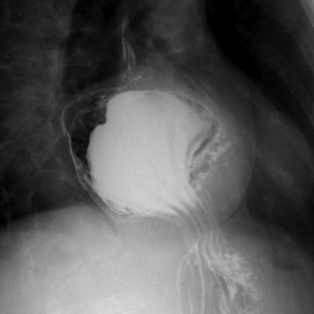
Barium swallow demonstrates a very large hiatal hernia. The stomach is normally below the heart in the abdomen. This contrast study demonstrates the abnormal location of the stomach. Notice how there is pooling of contrast in a ball located in the middle of the chest behind the heart.
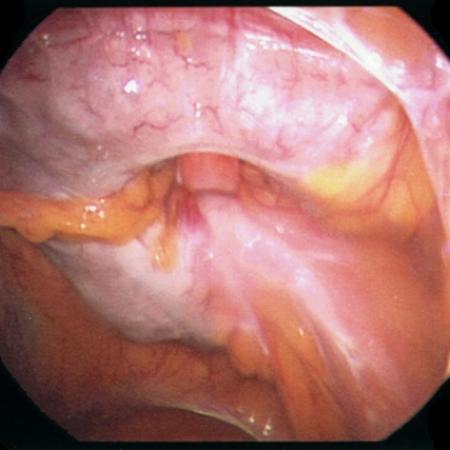
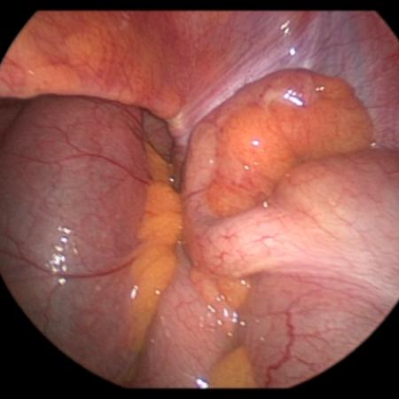
The Triangle of Calot is demonstrated during laparoscopic cholecystectomy in this photo. Although laparoscopic cholecystectomy is a very common operation, there are accepted complications. Complications are minimized by excellent exposure. The gallbladder is being adequately retracted allowing the…