
Robotic Removal of a Lobe of the Lung (Lobectomy)
The use of surgical procedures to treat lung cancer depends a great deal on the type of cancer, its stage (progression of the disease), location of the tumor(s) and the general health of the person.
There are two common types of lung cancer, small-cell lung cancer (SCLC) and non-small cell lung cancer (NSCLC). Small cell lung cancer is notorious for spreading early in the disease, which usually indicates that radiation or chemotherapy treatment is the most effective approach. With non-small cell lung cancer the spread of tumors is generally slower and more localized, which makes this type of cancer a candidate for surgery. Since non-small cell lung cancer accounts for more than 80% of all lung cancer cases, surgery is the most common treatment.
Robotic Lobectomy is Truly Minimally Invasive

Most lung cancers are carcinomas; malignancies that begin in epithelial cells (think surface cells such as skin). In this case these are the cells that line the interior of the lungs. From these cells the growth of cancer usually leads to tumors, clumps of cancerous cells, which with the cancer type NSCLC can become quite large and prominent. Confirmation of these tumors through chest radiology (X-ray) or computer assisted tomography (CT scan) and biopsy usually includes a diagnosis of the clinical stage of the cancer. Clinical stages for NSCLC, which are determined before treatment, are rated from 1A (best prognosis) to IV (worst prognosis) and depend on the spread and condition of the cancer. The stage determination is usually crucial to potential treatment. For example, a Stage III lung cancer might first be treated with radiation or chemotherapy to reduce tumor size before surgery. This might not be the case with a Stage II cancer.
Along with the type and stage of the lung cancer, location of the tumor – where it occurs in the lung and the degree to which it penetrates nearby areas – may determine the best approach for surgery. There are, of course, two lungs – right and left – the left being nearest the heart. Each lung is divided into lobes, naturally occurring segments. The right lung has three lobes, the left has two lobes. The position of the tumor(s) within the lobes is an important factor in determining the surgical procedure.
Why is it sometimes necessary to remove part of a lung?
Depending on the stage, size and location of tumor(s) within a lung, the most common surgical procedure is to remove an entire lobe, in effect, removing part of a lung. Experience has shown that lobe removal (a lobectomy) is more likely to encompass the cancer even if the tumor itself is located well within the lobe. Removing all the cancer is the goal, leaving behind even a few cancerous cells can lead to regeneration. Unfortunately, the natural division of the lungs into lobes doesn’t provide a true barrier to cancer. The ultimate danger is the beginning of metastasis, or spread of cancer from one lobe to another, or to other parts of the body.
There are principally three approaches to lobectomy: Open, VATS and minimally invasive robotic surgery.
With an open lobectomy an incision is made on the side of the chest nearly the length of the lung area. This is called a thoracotomy. The large incision, exposing the lung for the lobectomy procedure, provides a clear view for the surgeon and an open path for removing tissue from the body. Typically this procedure requires general anesthesia and a breathing tube (endotracheal tube) connected to a ventilator machine. Obviously this approach is more traumatic for the patient, but may be the only practical method if the tumor is very large or the spread of cancer is somewhat uncertain.
Combining technology for the best approach

The Video-Assisted Thoracoscopic Surgery (VATS) approach, which is perhaps the most commonly used, consists of at least one relatively small incision for insertion of surgical instruments or to remove tissue, and another incision to insert a camera (endoscope). The surgeon uses the video image from the camera inside the body, usually displayed in a monitor just above the operating table, to see the surgical field (area of work). Though less invasive than a full open thoracotomy, the VATS approach often uses two or more incisions, often called ports or portals, including one to allow a ‘hand assist’ – the insertion of a human hand. Combined, these ports can expose almost as much area as an open thoracotomy. The procedure also requires a surgical assistant to manipulate the interior camera for the primary surgeon, who is not able to view what the surgical instruments are doing without the camera’s images. Coordination in this procedure can be tricky.
The third approach is robotic assisted minimally invasive surgery, which uses very small incisions, usually about the size of a dime except possibly for the incision (or port) to remove tissue. Instead of manually performing the procedure, the surgeon controls robotic devices (arms with instruments).
Which of these approaches to lobectomy is recommended will depend on the factors mentioned: size of tumor, location and stage, plus current lung function and general state of health. As might be expected a patient who is not in good general health or whose lung capacity is already greatly reduced is less of a candidate for open lobectomy – or even for a lobectomy at all.
What are the advantages of robotic minimally invasive surgery?
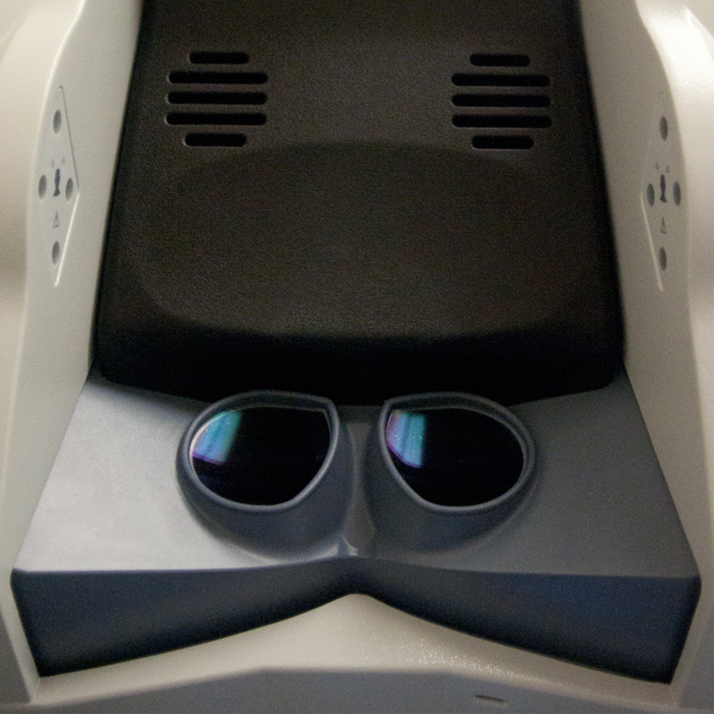
A robotic lobectomy does not mean a robot removes the lobe. The primary surgeon controls the procedure through a robotic device called the da Vinci Surgery System. In this system the surgeon sits at a sophisticated electronic console, views the procedure through a special three-dimensional video image, and controls the action of robotic arms and instruments with both hands and feet. The da Vinci system assists or augments the movements of the surgeon. It makes no decisions, performs no movements of its own, nor is it programmed to perform the procedure.
Specialists working together
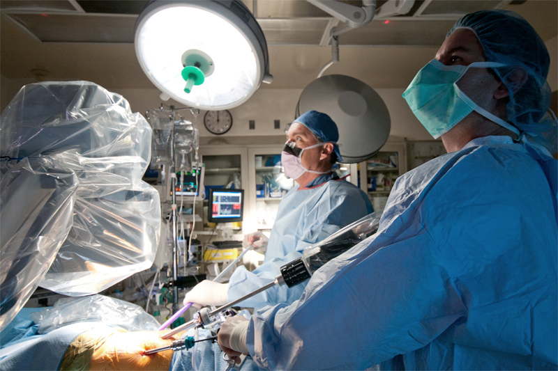
While the surgeon’s console is in the operating room and the patient is visible, the working ends of the system are four robotic arms mounted on a supporting cart that is placed at the side of the operating table and the patient. At the end of each arm a surgical instrument can be mounted, such as surgical knife or clamp. All of the instruments are miniaturized and connected to motor driven joints (servos) that provide very fine control in a very small space. In fact, the da Vinci system can provide flexibility and articulation, such as ‘wristing,’ at a level of precision beyond that of the human hand.
How the lung is accessed matters a great deal. A thoracotomy has advantages for the surgeon, but when it comes to recovery it is known to be among the most painful forms of surgery for the patient. In fact, the pain can make it difficult for the patient to breathe normally, which can lead to pneumonia or collapse of air respiration (atelectasis). This can even be a problem with the VATS procedure.
In contrast, a robotic minimally invasive surgery leaves only tiny incisions, which heal more quickly and with less pain. Internally, the da Vinci system presents a powerful combination: One part is an exceptionally clear view. The camera, which is controlled by the surgeon, produces computer-enhanced stereoscopic (3-D) images that can compensate for movement and the usually murky interior of the human body. The fine motor control of the robotic instruments makes it possible to perform certain actions, such as lymph node dissection, one of the typical surgical procedures for the lobectomy, with greater precision and speed than if it were done by hand. The end result is that while a robotic lobectomy usually requires at least as much time as an open procedure, it is generally less traumatic to the patient.